cardiac muscle tissue under microscope
Terms in this set 15 Mesothelium simple squamous Eggs or. Cardiac muscle tissue is a type of muscle tissue found only in the heart.
 |
| Cardiac Muscle |
Which of the following is true about cardiac muscle cells.
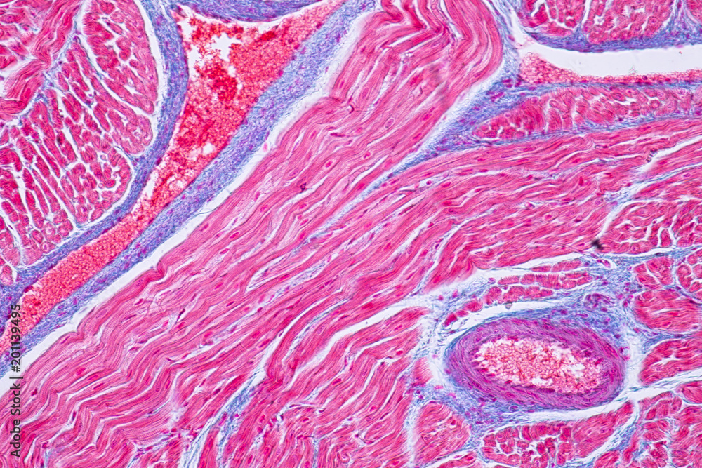
. The bundles are branched like a tree but. Describe the unique characteristics you. These muscles cannot move. Cardiac muscle is one of two muscle tissue types that is striated.
Tissues under a microscope. The bundles are branched like a tree but connected at both ends. Under the light microscope muscle cells appear striated with many nuclei squeezed along the membranes. What does cardiac muscle look like under the microscope.
Sarcomeres appear under the microscope as striations with alternating dark and light. Has intercalated discs that allow for communication between cardiac muscle cells What kind of muscle tissue is this. Where we can see cardiac muscle. Cardiac muscle Cardiac muscle tissue like skeletal muscle tissue looks striated or striped.
Unlike the other type of striated muscle tissue cardiac muscle tissue possesses intercalated disks that sit in. Unlike skeletal muscle tissue the contraction. Cardiac muscle tissue like skeletal muscle tissue looks striated or striped. Cardiac muscle is a special type of involuntary muscle present in the wall of the heart.
How will you identify columnar epithelial cells. These cells are also known as myocardium in vertebrates. Where is this tissue located. 2 MARK A student was observing a slide under a.
The bundles are branched like a tree but connected at both ends. It appears striated striped under a microscope due to the presence of sarcomere units that are. Location of cardiac tissue You will find the cardiac muscle in animals hearts and the larger vessels attached to the heart. It pumps blood through the body and is under involuntary control.
A sarcomere is the basic unit of muscle tissue in both cardiac and skeletal muscle. The specialized intercellular junctions found in cardiac muscle are a. Skeletal muscle tissue is arranged in bundles surrounded by connective tissue. Human cardiac muscle captured under the RB30 microscope at 40x magnification with a 5mp microscope camera.
What is cardiac muscles function. Blood vessels attached to the heart The function of cardiac muscle The cardiac muscle is involuntary and it is stimulated to contract by a mechanism similar to the skeletal muscle. Unlike skeletal muscle tissue. Cardiac muscle tissue like skeletal muscle tissue looks striated or striped.
Muscle tissue - cardiac Note. While observing skeletal muscle tissue under the microscope you note that it appears striated. Cardiac muscle tissue is. You tell your lab partner this is because.
 |
| Histology Human Cardiac Muscle Under Microscope Stock Photo 1120607303 Shutterstock |
 |
| Histology Of Human Cardiac Muscle Under Microscope View Stock Photo Picture And Royalty Free Image Image 74959415 |
 |
| H E Stained Cardiac Muscle Tissue Galleries Nikon Instruments Inc |
 |
| Histology Of Human Cardiac Muscle Under Microscope View For Education Stock Photo Image Of Contraction Medicine 183352432 |
 |
| Cardiac Muscle |

Posting Komentar untuk "cardiac muscle tissue under microscope"